
How AI May Support Faster Leukemia Investigations
A model can process an image quickly while the complete investigation still takes time. Understanding where AI may help requires examining the work before and after the model runs.
Identify the task first
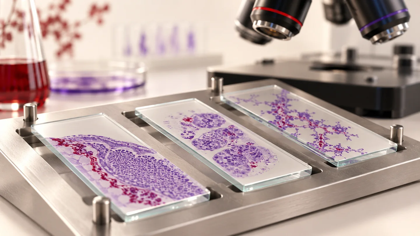
Digital-image research includes locating cells, assigning image labels and combining information across a specimen. The SGLNet study, for example, investigates a spatially guided approach to detecting leukemia-related cells in bone marrow images. The published abstract reports task-specific detection results, not a measured reduction in the time patients wait for a diagnosis. [1]
This difference matters when describing speed. Model execution is only one step in a wider process.
Measure the whole workflow
For a useful assessment, we recommend recording sample preparation, image acquisition, quality checks, processing, human review and reporting separately. That makes it possible to identify where a tool changes the work and where it introduces additional review.
Teams should also examine unsuccessful runs. A fast result on an ideal image does not explain what happens when a file is unreadable or when the output needs correction.
Preserve the evidence behind the result
A research record should connect the input image, processing version, output and reviewer action. Without these details, faster output may simply create a faster route to an uncheckable conclusion.
In our view, useful progress combines efficiency with clarity: a reviewer should understand what was analyzed, what the result represents and where further evidence is needed.
CellSight's role
CellSight's working first version supports connected blood-cell image research workflows. The team's internal functional testing does not establish a clinical turnaround-time benefit, and this article reports no CellSight speed or accuracy figure.
The platform is not for diagnosis or clinical decision-making. The appropriate research question is whether a defined workflow improves under measured conditions, rather than whether AI is faster in general.
References
- 1. High-efficiency spatially guided learning network for lymphoblastic leukemia detection in bone marrow microscopy images. Mei et al., Computers in Biology and Medicine. 2025-08-02 online; September 2025 issue. Accessed 2026-09-14.
Related Insights
AI and the Future of Leukemia Research
Explore how blood-cell imaging, AI and connected evidence can support leukemia research, and why meaningful evaluation extends beyond model accuracy.
By CellSight Editorial Team · September 13, 2026 · 2 min read
Read article
AI in Leukemia Microscopy: A Research Review
Review selected AI studies in leukemia microscopy, covering hybrid models, cell detection and case-level learning, with practical limits on comparing results.
By CellSight Editorial Team · September 13, 2026 · 3 min read
Read article
Advances in Leukemia Detection and Monitoring
Understand how molecular testing, flow cytometry, liquid-biopsy research and digital imaging contribute different evidence to leukemia investigations.
By CellSight Editorial Team · September 13, 2026 · 2 min read
Read article